Breast Screening
Breast Cancer Screening is a proactive step to detect breast cancer early.
Breast self-examination, clinical breast examination, and mammographic screenings are possible methods.
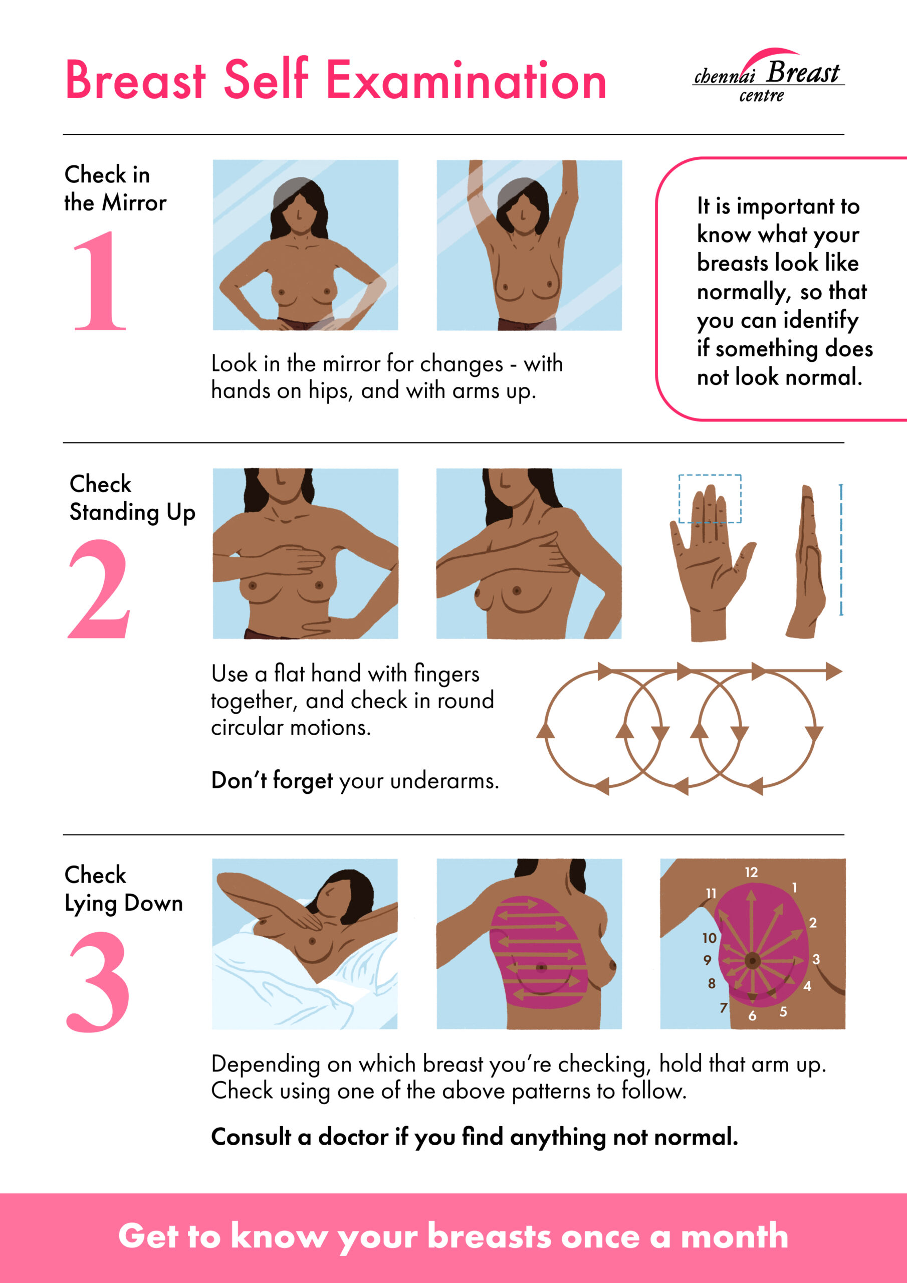
Breast Self Examination
Breast is a very dynamic organ and keeps changing with menstrual cycles, pregnancy, lactation, age, weight gain and weight loss. Being aware of these normal changes and the texture and feel of one’s own breasts helps in noticing changes early.
Timing and Technique for Breast Self-Examination
Why it helps:
- Regular checks help you become familiar with how your breasts normally look and feel.
- Any new or unusual changes can be detected early and evaluated by a doctor.
When to check:
- For menstruating women, it is best to examine the breasts after periods, as they can be tender and lumpy just before menstruation.
- Women who have gone through menopause can perform the check any day of the month, ideally once a month.
How to check:
- Use your hands and eyes to touch, look, and feel the breasts regularly.
- Doing this in the shower with soapy hands can make it easier to notice changes.
Mammographic Screening
Breast screening with mammography is being debated across the world with regards to its safety and overall survival benefit. There is no specific mammographic screening policy in India as per our health authorities. We have adapted the guidelines recommended by the UK guidelines and the American College of Radiologists and Cancer Society.
We recommend mammographic breast screening every 2 years for women over 40 years. Women with a higher risk (ie) strong family history of breast and ovarian cancer are recommended to have annual mammograms. Additionally, Breast MRI will also be required in women who have inherited breast cancer gene mutations.
When abnormalities are detected on screening, further diagnostic workup that includes a breast Ultrasound and image-guided (Stereo or Ultrasound-guided) breast biopsies will be performed.
Is mammography painful?
Undergoing a mammogram is uncomfortable for some women. The breast compression that is done during a mammogram is slightly uncomfortable. When a screening mammogram is done as part of a routine check up, it is best done a week after completion of periods to avoid discomfort.
There is very little pain associated with undergoing a mammography. It takes only a few moments and the discomfort is over soon.
Is it safe to have mammograms routinely?
Mammograms expose the breast to some radiation. Our tomo-synthesis machine minimises this radiation exposure by synthesizing 2D images with a special software from 3D images without losing resolution. It is safe to have a mammogram every two years starting at the age of 40.
Breast Cancer Diagnosis
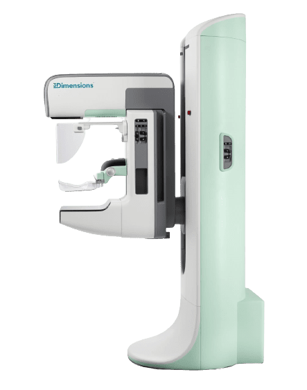
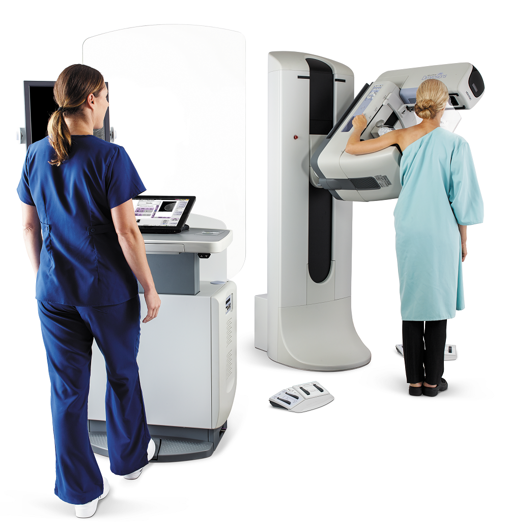
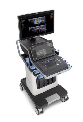
Chennai Breast Centre is equipped with the fastest, highest resolution breast tomosynthesis mammography system with superior-performing low dose 3D tomo exam with more accuracy. This technology is particularly useful in women with dense breasts. Our compassionate and skilled mammographers ensure that the screening and diagnostic process is as comfortable, reassuring and stress free.
- 3D HD Tomo Mammogram
- Bard Core Needle Biopsy
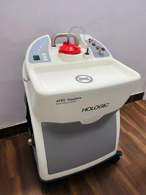
- Atec Suros Biopsy System
- Upright Stereo Biopsy System
- Supersonic High - Resolution Ultrasound
Diagnostic Mammogram
A diagnostic mammogram is performed for any symptom in the breast. The diagnostic mammogram is usually accompanied by a breast Ultrasound. Further tests like Ultrasound guided or stereotactic biopsy will be performed based on the findings on Mammogram and Breast Ultrasound.
3D HD Tomosynthesis Mammogram
Chennai Breast Centre is equipped with the latest and the best in mammographic technology.
- The fastest, highest resolution breast tomosynthesis mammography system ever in India.
- The highest resolution breast tomosynthesis exam with greater comfort.
- Clinically proven to deliver a more comfortable mammogram.
- Superior-performing low radiation dose 3D exam with more accuracy.
- Designed for greater clinical confidence.
- Designed to enhance the technologist work-flow.
What makes Chennai Breast Centre a trusted place for your mammogram?
- State of the art technology.
- Expert Interpretation & Consultation
- Mammogram, breast ultrasound, and a breast specialist’s opinion — all conveniently available in a single appointment.
- Tests and reports along with Doctor Consultation immediately.
- Women doctors and staff , delivering the best breast cancer care with skill, strength, and heartfelt empathy.
- A clear diagnosis with an appropriate recommendation.
- Images are archived for comparison at a later date. There is no risk of losing films and reports.
- Technologists are well trained to do the procedure with skill and sensitivity for optimal diagnosis, better patient experience and minimal pain.
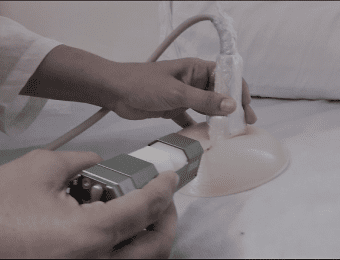
Breast Ultrasound
Generally, a breast ultrasound also accompanies a diagnostic mammography. Breast ultrasound is done in patients less than 40 years or as an additional modality along with mammogram to characterize lesions better. It is particularly useful to complement mammograms in women with dense breasts.
In an ultrasound, sound waves are transmitted to reflect a visual image of the interior of the breast. The process is absolutely painless and does not involve any radiation. The fluid-filled cysts are rapidly and accurately distinguished from the solid ones. The size, shape, location and the extent of the abnormality are clearly documented. Ultrasound examination is used to gain valuable details about the nature of the lesion, extent and guide biopsy/ intervention when it becomes necessary.
Chennai Breast Centre is equipped with high-resolution ultrasound machines, supported by skilled technologists and doctors who can diagnose even complex breast conditions.
Ultrasound Guided Core Needle Biopsy
- If any abnormality is detected on imaging, then the next step for diagnosis is an Ultrasound guided core needle biopsy.
- Ultrasound guided core needle biopsy is performed under local anaesthesia. A small piece of tissue is removed and sent for testing to the lab and results will be available in a few days.
- There is no hazard of spread of disease or cancer when a biopsy is done and this step is essential in planning further treatment and management.
Stereotactic and Vacuum Assisted Biopsy
Stereotactic biopsy is done when an abnormality is seen only on the mammogram. ( i.e) microcalcifications
Chennai Breast Centre is equipped with Affirm tomo biopsy unit with a reclining stereo chair. With the Affirm® 3D™ upright tomo biopsy unit, even the lesions that are visible only with tomosynthesis imaging are also clearly localised and biopsied accurately.
Our centre is equipped with the Suros ATEC Vacuum-assisted breast biopsy technology. A biopsy procedure with ATEC involves:
- A one-time needle insertion under local anesthesia
- No stitches
- No hospitalization
- No general anesthesia
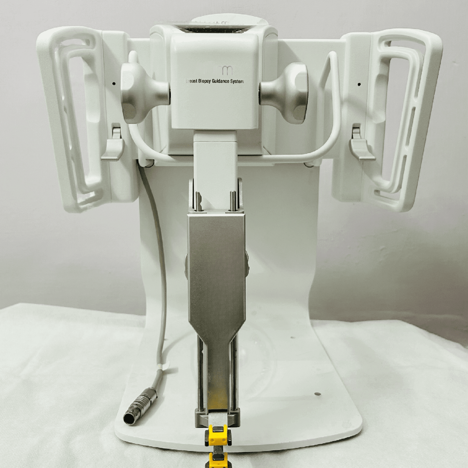
This ensures high-quality samples for a definitive diagnosis and allows the patient to return to normal activities immediately.
Once the biopsy reports are available, the next steps in treatment will be planned based on those results.